World’s First Brain Stimulation Treatment for Neurological Diseases Uses Ultrasound Technology
|
By MedImaging International staff writers Posted on 19 Dec 2023 |

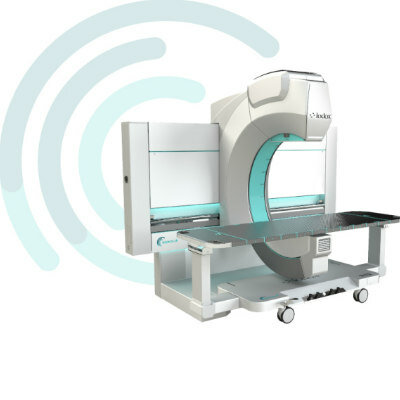
The effectiveness of drug treatments for individuals with neurological diseases like Parkinson's disease and epilepsy often diminishes over time, necessitating alternative therapeutic approaches. Movement symptoms in such conditions are typically the result of disorganized neural signals in the brain areas governing movement. Deep brain stimulation, a technique that disrupts these erratic signals, has shown promise in managing tremors and other movement-related symptoms. It also holds the potential for controlling, and possibly even preventing, seizures in epilepsy patients. Now, researchers are exploring a groundbreaking brain stimulation treatment that employs ultrasound, delivered through a device located inside a brain blood vessel, to address symptoms of neurological disorders.
A group of biomedical engineers at The University of Melbourne (Victoria, Australia), along with their colleagues, are developing ‘endovascular focused ultrasound’ technology, which could substantially enhance the lives of those with neurological diseases. Currently, electrical brain stimulation is a treatment option, but it requires invasive brain surgery to implant electrodes and often lacks precision in stimulating the correct brain regions. The accuracy of electrode placement is crucial; slight misplacements can lead to diminished efficacy and unintended side effects. The team's endovascular-focused ultrasound technology, anticipated to be available in over a decade, presents a safer alternative to existing treatments, avoiding the need for open brain surgery and utilizing ultrasound instead of electrical stimulation.
This innovative method involves inserting an array of tiny transducers permanently into a brain blood vessel, likely through the jugular vein, akin to current stent insertion practices in the heart and brain. These transducers would be connected to a chest-implanted device similar to a pacemaker, which would power and control the ultrasound stimulation. Thanks to advancements in nanofabrication technology, which is also used in computer chip manufacturing, researchers can produce extraordinarily small ultrasound transducers. These transducers are fine enough to fit inside a blood vessel, being narrower than human hair. When arranged in an array, they can precisely focus the ultrasound beam on a specific brain target, minimizing stimulation to surrounding tissue. The team aims for a successful initial demonstration of this novel brain stimulation method within a three-year project timeline and is hopeful that the first human trials could commence by 2030.
“We have demonstrated preliminary safety and efficacy of implanting neurotechnology within cortical blood vessels to enable people with paralysis to control external equipment with their minds,” said Professor Nicholas Opie from the University of Melbourne Department of Medicine. “Ultrasound is a new method of stimulating the brain and has the potential to provide transformative treatments for people with neurological disorders such as Parkinson’s, epilepsy and depression.”
Related Links:
The University of Melbourne
Latest Ultrasound News
- Deep Learning Advances Super-Resolution Ultrasound Imaging
- Novel Ultrasound-Launched Targeted Nanoparticle Eliminates Biofilm and Bacterial Infection
- AI-Guided Ultrasound System Enables Rapid Assessments of DVT
- Focused Ultrasound Technique Gets Quality Assurance Protocol
- AI-Guided Handheld Ultrasound System Helps Capture Diagnostic-Quality Cardiac Images
- Non-Invasive Ultrasound Imaging Device Diagnoses Risk of Chronic Kidney Disease
- Wearable Ultrasound Platform Paves Way for 24/7 Blood Pressure Monitoring On the Wrist
- Diagnostic Ultrasound Enhancing Agent to Improve Image Quality in Pediatric Heart Patients
- AI Detects COVID-19 in Lung Ultrasound Images
- New Ultrasound Technology to Revolutionize Respiratory Disease Diagnoses
- Dynamic Contrast-Enhanced Ultrasound Highly Useful For Interventions
- Ultrasensitive Broadband Transparent Ultrasound Transducer Enhances Medical Diagnosis
- Artificial Intelligence Detects Heart Defects in Newborns from Ultrasound Images
- Ultrasound Imaging Technology Allows Doctors to Watch Spinal Cord Activity during Surgery

- Shape-Shifting Ultrasound Stickers Detect Post-Surgical Complications
- Non-Invasive Ultrasound Technique Helps Identify Life-Changing Complications after Neck Surgery
Channels
Radiography
view channel
Novel Breast Imaging System Proves As Effective As Mammography
Breast cancer remains the most frequently diagnosed cancer among women. It is projected that one in eight women will be diagnosed with breast cancer during her lifetime, and one in 42 women who turn 50... Read more
AI Assistance Improves Breast-Cancer Screening by Reducing False Positives
Radiologists typically detect one case of cancer for every 200 mammograms reviewed. However, these evaluations often result in false positives, leading to unnecessary patient recalls for additional testing,... Read moreMRI
view channel
PET/MRI Improves Diagnostic Accuracy for Prostate Cancer Patients
The Prostate Imaging Reporting and Data System (PI-RADS) is a five-point scale to assess potential prostate cancer in MR images. PI-RADS category 3 which offers an unclear suggestion of clinically significant... Read more
Next Generation MR-Guided Focused Ultrasound Ushers In Future of Incisionless Neurosurgery
Essential tremor, often called familial, idiopathic, or benign tremor, leads to uncontrollable shaking that significantly affects a person’s life. When traditional medications do not alleviate symptoms,... Read more
Two-Part MRI Scan Detects Prostate Cancer More Quickly without Compromising Diagnostic Quality
Prostate cancer ranks as the most prevalent cancer among men. Over the last decade, the introduction of MRI scans has significantly transformed the diagnosis process, marking the most substantial advancement... Read moreNuclear Medicine
view channel
New SPECT/CT Technique Could Change Imaging Practices and Increase Patient Access
The development of lead-212 (212Pb)-PSMA–based targeted alpha therapy (TAT) is garnering significant interest in treating patients with metastatic castration-resistant prostate cancer. The imaging of 212Pb,... Read moreNew Radiotheranostic System Detects and Treats Ovarian Cancer Noninvasively
Ovarian cancer is the most lethal gynecological cancer, with less than a 30% five-year survival rate for those diagnosed in late stages. Despite surgery and platinum-based chemotherapy being the standard... Read more
AI System Automatically and Reliably Detects Cardiac Amyloidosis Using Scintigraphy Imaging
Cardiac amyloidosis, a condition characterized by the buildup of abnormal protein deposits (amyloids) in the heart muscle, severely affects heart function and can lead to heart failure or death without... Read moreGeneral/Advanced Imaging
view channel
New AI Method Captures Uncertainty in Medical Images
In the field of biomedicine, segmentation is the process of annotating pixels from an important structure in medical images, such as organs or cells. Artificial Intelligence (AI) models are utilized to... Read more.jpg)
CT Coronary Angiography Reduces Need for Invasive Tests to Diagnose Coronary Artery Disease
Coronary artery disease (CAD), one of the leading causes of death worldwide, involves the narrowing of coronary arteries due to atherosclerosis, resulting in insufficient blood flow to the heart muscle.... Read more
Novel Blood Test Could Reduce Need for PET Imaging of Patients with Alzheimer’s
Alzheimer's disease (AD), a condition marked by cognitive decline and the presence of beta-amyloid (Aβ) plaques and neurofibrillary tangles in the brain, poses diagnostic challenges. Amyloid positron emission... Read more.jpg)
CT-Based Deep Learning Algorithm Accurately Differentiates Benign From Malignant Vertebral Fractures
The rise in the aging population is expected to result in a corresponding increase in the prevalence of vertebral fractures which can cause back pain or neurologic compromise, leading to impaired function... Read moreImaging IT
view channel
New Google Cloud Medical Imaging Suite Makes Imaging Healthcare Data More Accessible
Medical imaging is a critical tool used to diagnose patients, and there are billions of medical images scanned globally each year. Imaging data accounts for about 90% of all healthcare data1 and, until... Read more
Global AI in Medical Diagnostics Market to Be Driven by Demand for Image Recognition in Radiology
The global artificial intelligence (AI) in medical diagnostics market is expanding with early disease detection being one of its key applications and image recognition becoming a compelling consumer proposition... Read moreIndustry News
view channel
Bayer and Google Partner on New AI Product for Radiologists
Medical imaging data comprises around 90% of all healthcare data, and it is a highly complex and rich clinical data modality and serves as a vital tool for diagnosing patients. Each year, billions of medical... Read more


