Hyperfine Demonstrates World’s First FDA-Cleared, Portable, Ultra-Low-Field, MR Brain Imaging System
|
By MedImaging International staff writers Posted on 27 Nov 2023 |

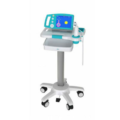
Hyperfine (Guilford, CT, USA) is showcasing the world’s first FDA-cleared, portable, ultra-low-field, magnetic resonance brain imaging system—the Swoop system—at RSNA 2023. The Swoop system which is capable of providing imaging at multiple points of care received initial U.S. Food and Drug Administration (FDA) clearance in 2020. It is a portable ultra-low-field MRI device for producing images that display the internal structure of the head where a full diagnostic examination is not clinically practical. When interpreted by a trained physician, these images provide information that can be useful in determining a diagnosis. Traditionally, access to costly, stationary, conventional MRI technology can be inconvenient or not available when needed most. With the portable, ultra-low-field Swoop system, Hyperfine, Inc. is redefining the neuroimaging workflow by bringing brain imaging to the patient’s bedside.
In addition, Hyperfine is presenting three scientific sessions highlighting ultra-low-field imaging data at RSNA 2023. These studies analyzed how portable MR brain imaging may assist physicians in the diagnosis and management of neurological conditions in critical care settings. The first session titled ‘Longitudinal Relaxivity Estimation of Gadolinium-based Contrast Agents’ is being presented by Sudarshan Ragunathan, Ph.D., and the second session titled ‘A Generative Adversarial Network Approach for Low-field to High-field Image Translation’ is being presented by Alfredo Lucas. The third session titled ‘Point-of-care Brain MRI: Update on Preliminary Results from a Single-center Retrospective Study in the Neurocritical Care Setting’ is being presented by Brian Gerard Yep, MD.
“At Hyperfine, Inc., we strive to push the boundaries of medical technology to make MR brain imaging more accessible, and our presence at RSNA 2023 underscores the promise of expanded use cases for portable MR brain imaging technology,” said Chip Truwit, MD, Hyperfine, Inc. vice president of scientific affairs. “We’ll continue to prioritize clinical application research for the Swoop system because expanding access to portable MR brain imaging is critical in improving how patient care is delivered.”
Related Links:
Hyperfine
Latest RSNA 2023 News
- Hologic Showcases Developments in Next-Generation AI Solutions
- Vieworks Unveils Groundbreaking Software Technologies Focusing on Safety and Efficiency
- Varex Imaging Showcases Nexus Line of Digital Image Acquisition Solutions
- Radcal Presents Accu-Gold TOUCH Series for X-Ray QA
- Bayer Presents Advancements in Radiology Pipeline and AI Innovations
- Philips Debuts World’s First Mobile MRI System With Helium-Free Operations
- Siemens Presents Groundbreaking Innovations Shaping the Future of Healthcare
- Alpinion Introduces New Paradigm in Ultrasound Diagnosis
- Fujifilm Unveils New Medical Imaging Innovations
- EDAN Instruments Demonstrates Flagship Models Offering Ease of Imaging in Lightweight Design
- GE HealthCare Showcases Innovative AI-Enabled Imaging Technology Solutions
- Mindray Showcases Latest Advancements in Radiology
- Samsung Ushers in New Era of Diagnostic Solutions
- United Imaging Showcases Pioneering Medical AI Solutions
- Subtle Medical Unveils Cutting-Edge Workflow Innovations
- Carestream Showcases Ideas that Clearly Work
Channels
Radiography
view channel
Novel Breast Imaging System Proves As Effective As Mammography
Breast cancer remains the most frequently diagnosed cancer among women. It is projected that one in eight women will be diagnosed with breast cancer during her lifetime, and one in 42 women who turn 50... Read more
AI Assistance Improves Breast-Cancer Screening by Reducing False Positives
Radiologists typically detect one case of cancer for every 200 mammograms reviewed. However, these evaluations often result in false positives, leading to unnecessary patient recalls for additional testing,... Read moreMRI
view channel
PET/MRI Improves Diagnostic Accuracy for Prostate Cancer Patients
The Prostate Imaging Reporting and Data System (PI-RADS) is a five-point scale to assess potential prostate cancer in MR images. PI-RADS category 3 which offers an unclear suggestion of clinically significant... Read more
Next Generation MR-Guided Focused Ultrasound Ushers In Future of Incisionless Neurosurgery
Essential tremor, often called familial, idiopathic, or benign tremor, leads to uncontrollable shaking that significantly affects a person’s life. When traditional medications do not alleviate symptoms,... Read more
Two-Part MRI Scan Detects Prostate Cancer More Quickly without Compromising Diagnostic Quality
Prostate cancer ranks as the most prevalent cancer among men. Over the last decade, the introduction of MRI scans has significantly transformed the diagnosis process, marking the most substantial advancement... Read moreUltrasound
view channel
Deep Learning Advances Super-Resolution Ultrasound Imaging
Ultrasound localization microscopy (ULM) is an advanced imaging technique that offers high-resolution visualization of microvascular structures. It employs microbubbles, FDA-approved contrast agents, injected... Read more
Novel Ultrasound-Launched Targeted Nanoparticle Eliminates Biofilm and Bacterial Infection
Biofilms, formed by bacteria aggregating into dense communities for protection against harsh environmental conditions, are a significant contributor to various infectious diseases. Biofilms frequently... Read moreNuclear Medicine
view channel
New SPECT/CT Technique Could Change Imaging Practices and Increase Patient Access
The development of lead-212 (212Pb)-PSMA–based targeted alpha therapy (TAT) is garnering significant interest in treating patients with metastatic castration-resistant prostate cancer. The imaging of 212Pb,... Read moreNew Radiotheranostic System Detects and Treats Ovarian Cancer Noninvasively
Ovarian cancer is the most lethal gynecological cancer, with less than a 30% five-year survival rate for those diagnosed in late stages. Despite surgery and platinum-based chemotherapy being the standard... Read more
AI System Automatically and Reliably Detects Cardiac Amyloidosis Using Scintigraphy Imaging
Cardiac amyloidosis, a condition characterized by the buildup of abnormal protein deposits (amyloids) in the heart muscle, severely affects heart function and can lead to heart failure or death without... Read moreGeneral/Advanced Imaging
view channel
New AI Method Captures Uncertainty in Medical Images
In the field of biomedicine, segmentation is the process of annotating pixels from an important structure in medical images, such as organs or cells. Artificial Intelligence (AI) models are utilized to... Read more.jpg)
CT Coronary Angiography Reduces Need for Invasive Tests to Diagnose Coronary Artery Disease
Coronary artery disease (CAD), one of the leading causes of death worldwide, involves the narrowing of coronary arteries due to atherosclerosis, resulting in insufficient blood flow to the heart muscle.... Read more
Novel Blood Test Could Reduce Need for PET Imaging of Patients with Alzheimer’s
Alzheimer's disease (AD), a condition marked by cognitive decline and the presence of beta-amyloid (Aβ) plaques and neurofibrillary tangles in the brain, poses diagnostic challenges. Amyloid positron emission... Read more.jpg)
CT-Based Deep Learning Algorithm Accurately Differentiates Benign From Malignant Vertebral Fractures
The rise in the aging population is expected to result in a corresponding increase in the prevalence of vertebral fractures which can cause back pain or neurologic compromise, leading to impaired function... Read moreImaging IT
view channel
New Google Cloud Medical Imaging Suite Makes Imaging Healthcare Data More Accessible
Medical imaging is a critical tool used to diagnose patients, and there are billions of medical images scanned globally each year. Imaging data accounts for about 90% of all healthcare data1 and, until... Read more
Global AI in Medical Diagnostics Market to Be Driven by Demand for Image Recognition in Radiology
The global artificial intelligence (AI) in medical diagnostics market is expanding with early disease detection being one of its key applications and image recognition becoming a compelling consumer proposition... Read moreIndustry News
view channel
Bayer and Google Partner on New AI Product for Radiologists
Medical imaging data comprises around 90% of all healthcare data, and it is a highly complex and rich clinical data modality and serves as a vital tool for diagnosing patients. Each year, billions of medical... Read more


