Deep Learning Enables Fast 3D Brain MRI at 0.055 Tesla
|
By MedImaging International staff writers Posted on 27 Sep 2023 |

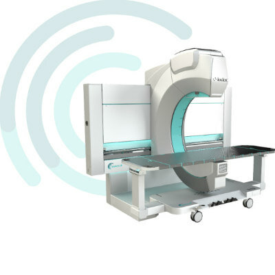
In the past few years, there has been significant work on developing portable ultralow-field magnetic resonance imaging (MRI) machines. These are designed to be affordable, easy to use without special shielding, and good for point-of-care settings. However, their image quality has not been up to the mark, and scans also take a long time. Deep learning is changing the game and has been effective in various high-field MR image reconstruction tasks, including artifact, denoising, and reconstruction. Now, researchers have proposed a fast acquisition and deep learning reconstruction framework to speed up brain MRI at 0.055 tesla.
In a new study aiming to advance brain ultralow-field MRI for speed and quality using deep learning, researchers at The University of Hong Kong (Hong Kong, PRC) successfully achieved fast 3D brain MRI at 0.055 T. This was done by directly combining the accelerated MR data acquisition and the deep learning 3D image formation that reconstructs images from incomplete 3D k-space data with super-resolution. The acquisition comprises a single average 3D encoding with 2D partial Fourier sampling, cutting down the scan time of T1- and T2-weighted imaging protocols to 2.5 and 3.2 minutes, respectively.
The 3D deep learning system takes advantage of the homogeneous brain anatomy available from high-field MRIs to enhance image quality. It minimizes errors and noise while boosting the spatial resolution to a synthetic 1.5-mm isotropic resolution. The new approach successfully overcomes the limitations of low signal strength, allowing for the detailed reconstruction of fine anatomical features. These features are consistent both within individual subjects and across different imaging protocols. This new method opens the door to fast and high-quality whole-brain MRIs at a magnetic field strength of 0.055 tesla, offering a variety of potential applications in biomedicine.
Related Links:
The University of Hong Kong
Latest MRI News
- PET/MRI Improves Diagnostic Accuracy for Prostate Cancer Patients
- Next Generation MR-Guided Focused Ultrasound Ushers In Future of Incisionless Neurosurgery
- Two-Part MRI Scan Detects Prostate Cancer More Quickly without Compromising Diagnostic Quality
- World’s Most Powerful MRI Machine Images Living Brain with Unrivaled Clarity
- New Whole-Body Imaging Technology Makes It Possible to View Inflammation on MRI Scan
- Combining Prostate MRI with Blood Test Can Avoid Unnecessary Prostate Biopsies
- New Treatment Combines MRI and Ultrasound to Control Prostate Cancer without Serious Side Effects
- MRI Improves Diagnosis and Treatment of Prostate Cancer
- Combined PET-MRI Scan Improves Treatment for Early Breast Cancer Patients
- 4D MRI Could Improve Clinical Assessment of Heart Blood Flow Abnormalities
- MRI-Guided Focused Ultrasound Therapy Shows Promise in Treating Prostate Cancer
- AI-Based MRI Tool Outperforms Current Brain Tumor Diagnosis Methods
- DW-MRI Lights up Small Ovarian Lesions like Light Bulbs
- Abbreviated Breast MRI Effective for High-Risk Screening without Compromising Diagnostic Accuracy
- New MRI Method Detects Alzheimer’s Earlier in People without Clinical Signs
- MRI Monitoring Reduces Mortality in Women at High Risk of BRCA1 Breast Cancer
Channels
Radiography
view channel
Novel Breast Imaging System Proves As Effective As Mammography
Breast cancer remains the most frequently diagnosed cancer among women. It is projected that one in eight women will be diagnosed with breast cancer during her lifetime, and one in 42 women who turn 50... Read more
AI Assistance Improves Breast-Cancer Screening by Reducing False Positives
Radiologists typically detect one case of cancer for every 200 mammograms reviewed. However, these evaluations often result in false positives, leading to unnecessary patient recalls for additional testing,... Read moreUltrasound
view channel
Deep Learning Advances Super-Resolution Ultrasound Imaging
Ultrasound localization microscopy (ULM) is an advanced imaging technique that offers high-resolution visualization of microvascular structures. It employs microbubbles, FDA-approved contrast agents, injected... Read more
Novel Ultrasound-Launched Targeted Nanoparticle Eliminates Biofilm and Bacterial Infection
Biofilms, formed by bacteria aggregating into dense communities for protection against harsh environmental conditions, are a significant contributor to various infectious diseases. Biofilms frequently... Read moreNuclear Medicine
view channel
New SPECT/CT Technique Could Change Imaging Practices and Increase Patient Access
The development of lead-212 (212Pb)-PSMA–based targeted alpha therapy (TAT) is garnering significant interest in treating patients with metastatic castration-resistant prostate cancer. The imaging of 212Pb,... Read moreNew Radiotheranostic System Detects and Treats Ovarian Cancer Noninvasively
Ovarian cancer is the most lethal gynecological cancer, with less than a 30% five-year survival rate for those diagnosed in late stages. Despite surgery and platinum-based chemotherapy being the standard... Read more
AI System Automatically and Reliably Detects Cardiac Amyloidosis Using Scintigraphy Imaging
Cardiac amyloidosis, a condition characterized by the buildup of abnormal protein deposits (amyloids) in the heart muscle, severely affects heart function and can lead to heart failure or death without... Read moreGeneral/Advanced Imaging
view channel
New AI Method Captures Uncertainty in Medical Images
In the field of biomedicine, segmentation is the process of annotating pixels from an important structure in medical images, such as organs or cells. Artificial Intelligence (AI) models are utilized to... Read more.jpg)
CT Coronary Angiography Reduces Need for Invasive Tests to Diagnose Coronary Artery Disease
Coronary artery disease (CAD), one of the leading causes of death worldwide, involves the narrowing of coronary arteries due to atherosclerosis, resulting in insufficient blood flow to the heart muscle.... Read more
Novel Blood Test Could Reduce Need for PET Imaging of Patients with Alzheimer’s
Alzheimer's disease (AD), a condition marked by cognitive decline and the presence of beta-amyloid (Aβ) plaques and neurofibrillary tangles in the brain, poses diagnostic challenges. Amyloid positron emission... Read more.jpg)
CT-Based Deep Learning Algorithm Accurately Differentiates Benign From Malignant Vertebral Fractures
The rise in the aging population is expected to result in a corresponding increase in the prevalence of vertebral fractures which can cause back pain or neurologic compromise, leading to impaired function... Read moreImaging IT
view channel
New Google Cloud Medical Imaging Suite Makes Imaging Healthcare Data More Accessible
Medical imaging is a critical tool used to diagnose patients, and there are billions of medical images scanned globally each year. Imaging data accounts for about 90% of all healthcare data1 and, until... Read more
Global AI in Medical Diagnostics Market to Be Driven by Demand for Image Recognition in Radiology
The global artificial intelligence (AI) in medical diagnostics market is expanding with early disease detection being one of its key applications and image recognition becoming a compelling consumer proposition... Read moreIndustry News
view channel
Bayer and Google Partner on New AI Product for Radiologists
Medical imaging data comprises around 90% of all healthcare data, and it is a highly complex and rich clinical data modality and serves as a vital tool for diagnosing patients. Each year, billions of medical... Read more


