


New SPECT/CT Scanner to Help Providers Transition Away from SPECT-Only Systems
|
By MedImaging International staff writers Posted on 14 Jun 2022 |
|
")
Historically, single photon emission computed tomography/computed tomography (SPECT/CT) has not been accessible to all healthcare providers. Instead, many institutions continue to use SPECT-only gamma cameras, which cannot always accommodate the clinical needs of a modern molecular-imaging or radiology department. Now, a new SPECT/CT scanner with its optimized, low-dose imaging, intuitive workflow, and ability to fit into most existing SPECT rooms removes barriers to SPECT/CT adoption and helps providers to transition from SPECT-only and early-generation SPECT/CT systems.
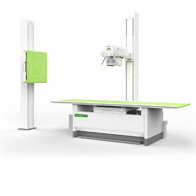
Siemens Healthineers (Erlangen, Germany) has launched Symbia Pro.specta, a SPECT/CT system with CE mark and Food and Drug Administration (FDA) clearance that has advanced SPECT and CT imaging technologies. Capabilities include a low-dose CT of up to 64 slices for impressive detail, automatic SPECT motion correction for additional image clarity, and an intuitive and automated workflow to guide the user through the entire decision-making process of the examination. Symbia Pro.specta is an all-purpose system that can be customized to accommodate a wide range of clinical exams, patient types, and department settings.
At the heart of Symbia Pro.specta is myExam Companion, which includes an intuitive user interface that eliminates the traditional manual and user-dependent SPECT/CT imaging workflow. myExam Companion provides automated tools to guide the user through every step of the exam’s decision-making process - from system and patient preparation to image acquisition and reconstruction to evaluation and post-processing. This enables departments to achieve consistent results quickly and more easily, regardless of the patient, procedure, or user experience level. Another major innovation that comes as standard is data-driven patient motion correction, which automatically corrects for patient movement in a SPECT exam with the click of a button, improving image quality without adding complexity. Additionally, data-driven respiratory motion correction for cardiac examinations is available as an option.
Designed for all SPECT/CT imaging applications, Symbia Pro.specta can be tailored as needed with specialized clinical tools for optimized imaging in cardiology, neurology, oncology, orthopedics, and more. Flexible detectors along with an accessible design facilitate imaging for a wide range of patient types - including pediatric, obese, and physically challenged patients - without compromising exam quality, patient comfort, or staff efficiency. The scanner also can be used for either stand-alone diagnostic CT or SPECT imaging, providing the user with the same intuitive interface for simpler operation.
Symbia Pro.specta features a minimum of 32 and a maximum of 64 CT slices, and it comes standard with a tin filter and CT iterative reconstruction for ultra-low patient and room dose. Its advanced quantification capabilities help the user determine the patient’s response to therapy. Capable of imaging at any energy level, the scanner is primed for imaging the high-energy isotopes increasingly used in theranostics, an approach that combines diagnostic and therapeutic agents to treat patients.
“Siemens Healthineers is proud to introduce the Symbia Pro.specta SPECT/CT scanner, which provides our customers with the ideal vehicle for transitioning from their SPECT-only and first-generation SPECT/CT cameras to a state-of-the-art SPECT/CT scanner that can perform a full spectrum of nuclear medicine examinations,” said Jim Williams, PhD, Head of Siemens Healthineers Molecular Imaging. “Symbia Pro.specta will help healthcare institutions overcome barriers to care by ensuring accessibility through its ease of use and ability to fit into existing SPECT rooms.”
Related Links:
Siemens Healthineers


Latest General/Advanced Imaging News
- New AI Method Captures Uncertainty in Medical Images
- CT Coronary Angiography Reduces Need for Invasive Tests to Diagnose Coronary Artery Disease
- Novel Blood Test Could Reduce Need for PET Imaging of Patients with Alzheimer’s
- CT-Based Deep Learning Algorithm Accurately Differentiates Benign From Malignant Vertebral Fractures
- Minimally Invasive Procedure Could Help Patients Avoid Thyroid Surgery
- Self-Driving Mobile C-Arm Reduces Imaging Time during Surgery
- AR Application Turns Medical Scans Into Holograms for Assistance in Surgical Planning
- Imaging Technology Provides Ground-Breaking New Approach for Diagnosing and Treating Bowel Cancer
- CT Coronary Calcium Scoring Predicts Heart Attacks and Strokes
- AI Model Detects 90% of Lymphatic Cancer Cases from PET and CT Images
- Breakthrough Technology Revolutionizes Breast Imaging
- State-Of-The-Art System Enhances Accuracy of Image-Guided Diagnostic and Interventional Procedures
- Catheter-Based Device with New Cardiovascular Imaging Approach Offers Unprecedented View of Dangerous Plaques
- AI Model Draws Maps to Accurately Identify Tumors and Diseases in Medical Images
- AI-Enabled CT System Provides More Accurate and Reliable Imaging Results
- Routine Chest CT Exams Can Identify Patients at Risk for Cardiovascular Disease
Channels
Radiography
view channel")
Novel Breast Imaging System Proves As Effective As Mammography
Breast cancer remains the most frequently diagnosed cancer among women. It is projected that one in eight women will be diagnosed with breast cancer during her lifetime, and one in 42 women who turn 50... Read more")
AI Assistance Improves Breast-Cancer Screening by Reducing False Positives
Radiologists typically detect one case of cancer for every 200 mammograms reviewed. However, these evaluations often result in false positives, leading to unnecessary patient recalls for additional testing,... Read more")
")
MRI
view channel")
PET/MRI Improves Diagnostic Accuracy for Prostate Cancer Patients
The Prostate Imaging Reporting and Data System (PI-RADS) is a five-point scale to assess potential prostate cancer in MR images. PI-RADS category 3 which offers an unclear suggestion of clinically significant... Read more")
Next Generation MR-Guided Focused Ultrasound Ushers In Future of Incisionless Neurosurgery
Essential tremor, often called familial, idiopathic, or benign tremor, leads to uncontrollable shaking that significantly affects a person’s life. When traditional medications do not alleviate symptoms,... Read more")
Two-Part MRI Scan Detects Prostate Cancer More Quickly without Compromising Diagnostic Quality
Prostate cancer ranks as the most prevalent cancer among men. Over the last decade, the introduction of MRI scans has significantly transformed the diagnosis process, marking the most substantial advancement... Read more of magnetic field vs. 1.5 and 3 T for conventional MRI machines in hospitals (Photo courtesy of CEA)")
Ultrasound
view channel")
Deep Learning Advances Super-Resolution Ultrasound Imaging
Ultrasound localization microscopy (ULM) is an advanced imaging technique that offers high-resolution visualization of microvascular structures. It employs microbubbles, FDA-approved contrast agents, injected... Read more")
Novel Ultrasound-Launched Targeted Nanoparticle Eliminates Biofilm and Bacterial Infection
Biofilms, formed by bacteria aggregating into dense communities for protection against harsh environmental conditions, are a significant contributor to various infectious diseases. Biofilms frequently... Read more")
")
Nuclear Medicine
view channel")
New SPECT/CT Technique Could Change Imaging Practices and Increase Patient Access
The development of lead-212 (212Pb)-PSMA–based targeted alpha therapy (TAT) is garnering significant interest in treating patients with metastatic castration-resistant prostate cancer. The imaging of 212Pb,... Read moreNew Radiotheranostic System Detects and Treats Ovarian Cancer Noninvasively
Ovarian cancer is the most lethal gynecological cancer, with less than a 30% five-year survival rate for those diagnosed in late stages. Despite surgery and platinum-based chemotherapy being the standard... Read more")
AI System Automatically and Reliably Detects Cardiac Amyloidosis Using Scintigraphy Imaging
Cardiac amyloidosis, a condition characterized by the buildup of abnormal protein deposits (amyloids) in the heart muscle, severely affects heart function and can lead to heart failure or death without... Read moreImaging IT
view channel")
New Google Cloud Medical Imaging Suite Makes Imaging Healthcare Data More Accessible
Medical imaging is a critical tool used to diagnose patients, and there are billions of medical images scanned globally each year. Imaging data accounts for about 90% of all healthcare data1 and, until... Read more
Global AI in Medical Diagnostics Market to Be Driven by Demand for Image Recognition in Radiology
The global artificial intelligence (AI) in medical diagnostics market is expanding with early disease detection being one of its key applications and image recognition becoming a compelling consumer proposition... Read more

Industry News
view channel")
Bayer and Google Partner on New AI Product for Radiologists
Medical imaging data comprises around 90% of all healthcare data, and it is a highly complex and rich clinical data modality and serves as a vital tool for diagnosing patients. Each year, billions of medical... Read more")
")
")