AI Suite Detects Most Common Chest X-Ray Findings
|
By MedImaging International staff writers Posted on 05 Oct 2021 |

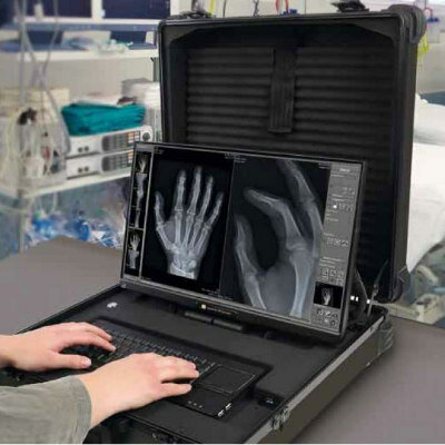
Image: Insight CXR detects abnormalities on a chest X-ray image (Photo courtesy of Lunit)
A new collaboration between Royal Philips (Amsterdam, The Netherlands) and Lunit (Seoul, South Korea) will make Lunit's AI software, the Insight CXR chest detection suite accessible to users of Philips' diagnostic X-ray solutions. CXR detects ten of the most common findings in a chest X-ray, incluindg small and subtle pulmonary nodules overlapped in the hilar shadow, ribs, heart, and diaphragm, enabling radiologists to reduce overlooked lung cancer cases, especially during regular check-ups.
Lunit Insight CXR is designed to instantly analyze of chest X-ray images by mapping radiological findings and displaying a scored calculation of actual existence. The algorithm shows a 97-99% accuracy rate in the detection of lung nodules, calcifications, consolidation, fibrosis, pneumothorax, pneumoperitoneum, cardiomegaly, pleural effusion, mediastinal widening, atelectasis, and tuberculosis. Data on the detected lesions is presented in the form of heatmaps and/or contour maps, with an abnormality score reflecting the AI’s calculation of the actual presence of the detected lesion.
“Radiology departments and their technologists are continually under pressure. They face high patient volumes, and every improvement in workflow can make a big impact,” said Daan van Manen, general manager for diagnostic X-ray at Philips. “Our partnership with Lunit to incorporate their diagnostic AI into our X-ray suite combines with a host of smart workflow features in the Philips radiography unified user interface (Eleva), across our digital radiography systems that enables a smooth and efficient, patient-focused workflow.”
“By partnering with Philips, one of the biggest medical device companies globally, our AI will be available to its significant global installed base. With the start of this partnership, we look forward to further expanding our collaboration to make data-driven medicine the new standard of care,” said Brandon Suh, CEO of Lunit. “Lunit will continue to build upon its current AI offering, making it better and better with time, and will continue to deliver best-in-class AI.”
Related Links:
Royal Philips
Lunit
Latest Radiography News
- Novel Breast Imaging System Proves As Effective As Mammography
- AI Assistance Improves Breast-Cancer Screening by Reducing False Positives
- AI Could Boost Clinical Adoption of Chest DDR
- 3D Mammography Almost Halves Breast Cancer Incidence between Two Screening Tests
- AI Model Predicts 5-Year Breast Cancer Risk from Mammograms
- Deep Learning Framework Detects Fractures in X-Ray Images With 99% Accuracy
- Direct AI-Based Medical X-Ray Imaging System a Paradigm-Shift from Conventional DR and CT
- Chest X-Ray AI Solution Automatically Identifies, Categorizes and Highlights Suspicious Areas
- AI Diagnoses Wrist Fractures As Well As Radiologists
- Annual Mammography Beginning At 40 Cuts Breast Cancer Mortality By 42%
- 3D Human GPS Powered By Light Paves Way for Radiation-Free Minimally-Invasive Surgery
- Novel AI Technology to Revolutionize Cancer Detection in Dense Breasts
- AI Solution Provides Radiologists with 'Second Pair' Of Eyes to Detect Breast Cancers
- AI Helps General Radiologists Achieve Specialist-Level Performance in Interpreting Mammograms
- Novel Imaging Technique Could Transform Breast Cancer Detection
- Computer Program Combines AI and Heat-Imaging Technology for Early Breast Cancer Detection
Channels
MRI
view channel
PET/MRI Improves Diagnostic Accuracy for Prostate Cancer Patients
The Prostate Imaging Reporting and Data System (PI-RADS) is a five-point scale to assess potential prostate cancer in MR images. PI-RADS category 3 which offers an unclear suggestion of clinically significant... Read more
Next Generation MR-Guided Focused Ultrasound Ushers In Future of Incisionless Neurosurgery
Essential tremor, often called familial, idiopathic, or benign tremor, leads to uncontrollable shaking that significantly affects a person’s life. When traditional medications do not alleviate symptoms,... Read more
Two-Part MRI Scan Detects Prostate Cancer More Quickly without Compromising Diagnostic Quality
Prostate cancer ranks as the most prevalent cancer among men. Over the last decade, the introduction of MRI scans has significantly transformed the diagnosis process, marking the most substantial advancement... Read moreUltrasound
view channel
Deep Learning Advances Super-Resolution Ultrasound Imaging
Ultrasound localization microscopy (ULM) is an advanced imaging technique that offers high-resolution visualization of microvascular structures. It employs microbubbles, FDA-approved contrast agents, injected... Read more
Novel Ultrasound-Launched Targeted Nanoparticle Eliminates Biofilm and Bacterial Infection
Biofilms, formed by bacteria aggregating into dense communities for protection against harsh environmental conditions, are a significant contributor to various infectious diseases. Biofilms frequently... Read moreNuclear Medicine
view channel
New SPECT/CT Technique Could Change Imaging Practices and Increase Patient Access
The development of lead-212 (212Pb)-PSMA–based targeted alpha therapy (TAT) is garnering significant interest in treating patients with metastatic castration-resistant prostate cancer. The imaging of 212Pb,... Read moreNew Radiotheranostic System Detects and Treats Ovarian Cancer Noninvasively
Ovarian cancer is the most lethal gynecological cancer, with less than a 30% five-year survival rate for those diagnosed in late stages. Despite surgery and platinum-based chemotherapy being the standard... Read more
AI System Automatically and Reliably Detects Cardiac Amyloidosis Using Scintigraphy Imaging
Cardiac amyloidosis, a condition characterized by the buildup of abnormal protein deposits (amyloids) in the heart muscle, severely affects heart function and can lead to heart failure or death without... Read moreGeneral/Advanced Imaging
view channel
New AI Method Captures Uncertainty in Medical Images
In the field of biomedicine, segmentation is the process of annotating pixels from an important structure in medical images, such as organs or cells. Artificial Intelligence (AI) models are utilized to... Read more.jpg)
CT Coronary Angiography Reduces Need for Invasive Tests to Diagnose Coronary Artery Disease
Coronary artery disease (CAD), one of the leading causes of death worldwide, involves the narrowing of coronary arteries due to atherosclerosis, resulting in insufficient blood flow to the heart muscle.... Read more
Novel Blood Test Could Reduce Need for PET Imaging of Patients with Alzheimer’s
Alzheimer's disease (AD), a condition marked by cognitive decline and the presence of beta-amyloid (Aβ) plaques and neurofibrillary tangles in the brain, poses diagnostic challenges. Amyloid positron emission... Read more.jpg)
CT-Based Deep Learning Algorithm Accurately Differentiates Benign From Malignant Vertebral Fractures
The rise in the aging population is expected to result in a corresponding increase in the prevalence of vertebral fractures which can cause back pain or neurologic compromise, leading to impaired function... Read moreImaging IT
view channel
New Google Cloud Medical Imaging Suite Makes Imaging Healthcare Data More Accessible
Medical imaging is a critical tool used to diagnose patients, and there are billions of medical images scanned globally each year. Imaging data accounts for about 90% of all healthcare data1 and, until... Read more
Global AI in Medical Diagnostics Market to Be Driven by Demand for Image Recognition in Radiology
The global artificial intelligence (AI) in medical diagnostics market is expanding with early disease detection being one of its key applications and image recognition becoming a compelling consumer proposition... Read moreIndustry News
view channel
Bayer and Google Partner on New AI Product for Radiologists
Medical imaging data comprises around 90% of all healthcare data, and it is a highly complex and rich clinical data modality and serves as a vital tool for diagnosing patients. Each year, billions of medical... Read more


