Power Assist Technology Positions X-Ray Tubes Effortlessly
|
By MedImaging International staff writers Posted on 14 Jan 2021 |

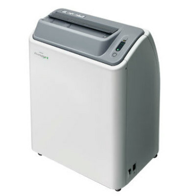
Image: The RADspeed Pro style edition with Power Glide (Photo courtesy of Shimadzu Medical Systems)
Brand-new power assist technology designed for high-end Shimadzu Medical Systems (SMS; Kyoto, Japan) general radiography systems provides an effortless operating assistance experience, improving radiographic examination environments. The assist-level can be selected among three levels on the X-ray tube support touch panel.
The technology, called Power Glide, is designed to instantaneously sense the amount of force applied by an operator during each operation, calculate the amount of assist-force required, and activate the motors accordingly to provide smooth and comfortable control when positioning the X-ray tube. As the forces applied to the handle during operation vary from person to person, the amount of power assist that each operator feels is optimal for operations, such as for large movements or detailed positioning adjustments, differs as well.
Power Glide is available in the SMS RADspeed Pro style edition, which provides up to 800 anatomical programs that can be registered; auto-positioning can move the position and angle of the X-ray tube to the programmed position. An auto-collimator selects a proper filter according to the program selected to optimize exposure dose and image quality. For dose management, the system can automatically calculate the dose area product (DAP) value from the X-ray conditions and display at each exposure. The LCD screen and illumination color can change according to the Bucky table or X-ray tube settings selected.
The ceiling X-ray tube support ensures wide-range vertical stroke of 1600 mm to examine the whole body (up to the lower extremities) in standing position. This support also rotates on the vertical and horizontal axis in addition to fixed positioning at any desired angle, enabling fast positioning at complex angles for orthopedic applications. Options include automatic imaging stitching; gridless scatter noise removal and improved image contrast; an image processing function that helps to confirm the absence of residual instruments, like gauze and needles, and reconfirmation of catheter tip location in PICC insertion procedures.
“Shimadzu has remained committed to developing innovative technologies that minimize radiation dose, improve image quality, and ensure excellent operability, in order to supply diagnostic imaging systems for supporting those working at the front lines of healthcare throughout the world,” said the company in a press release. “This new product can help increase the productivity of busy healthcare operations and improve the working environment of medical professionals, whether at small clinics or large hospitals, while maintaining the cumulative excellences offered by previous models.”
As general radiography examinations require fine operability to position the X-ray exposure area to within a few millimeters, it requires moving the entire assembly along ceiling rails from large distances, and as result the physical burden on operators is high. Reducing operating loads by improving system ergonomics enables a smoother workflow, and also shortens the time that patients must maintain a particular body position during examinations.
Latest Radiography News
- Novel Breast Imaging System Proves As Effective As Mammography
- AI Assistance Improves Breast-Cancer Screening by Reducing False Positives
- AI Could Boost Clinical Adoption of Chest DDR
- 3D Mammography Almost Halves Breast Cancer Incidence between Two Screening Tests
- AI Model Predicts 5-Year Breast Cancer Risk from Mammograms
- Deep Learning Framework Detects Fractures in X-Ray Images With 99% Accuracy
- Direct AI-Based Medical X-Ray Imaging System a Paradigm-Shift from Conventional DR and CT
- Chest X-Ray AI Solution Automatically Identifies, Categorizes and Highlights Suspicious Areas
- AI Diagnoses Wrist Fractures As Well As Radiologists
- Annual Mammography Beginning At 40 Cuts Breast Cancer Mortality By 42%
- 3D Human GPS Powered By Light Paves Way for Radiation-Free Minimally-Invasive Surgery
- Novel AI Technology to Revolutionize Cancer Detection in Dense Breasts
- AI Solution Provides Radiologists with 'Second Pair' Of Eyes to Detect Breast Cancers
- AI Helps General Radiologists Achieve Specialist-Level Performance in Interpreting Mammograms
- Novel Imaging Technique Could Transform Breast Cancer Detection
- Computer Program Combines AI and Heat-Imaging Technology for Early Breast Cancer Detection
Channels
MRI
view channel
PET/MRI Improves Diagnostic Accuracy for Prostate Cancer Patients
The Prostate Imaging Reporting and Data System (PI-RADS) is a five-point scale to assess potential prostate cancer in MR images. PI-RADS category 3 which offers an unclear suggestion of clinically significant... Read more
Next Generation MR-Guided Focused Ultrasound Ushers In Future of Incisionless Neurosurgery
Essential tremor, often called familial, idiopathic, or benign tremor, leads to uncontrollable shaking that significantly affects a person’s life. When traditional medications do not alleviate symptoms,... Read more
Two-Part MRI Scan Detects Prostate Cancer More Quickly without Compromising Diagnostic Quality
Prostate cancer ranks as the most prevalent cancer among men. Over the last decade, the introduction of MRI scans has significantly transformed the diagnosis process, marking the most substantial advancement... Read moreUltrasound
view channel
Deep Learning Advances Super-Resolution Ultrasound Imaging
Ultrasound localization microscopy (ULM) is an advanced imaging technique that offers high-resolution visualization of microvascular structures. It employs microbubbles, FDA-approved contrast agents, injected... Read more
Novel Ultrasound-Launched Targeted Nanoparticle Eliminates Biofilm and Bacterial Infection
Biofilms, formed by bacteria aggregating into dense communities for protection against harsh environmental conditions, are a significant contributor to various infectious diseases. Biofilms frequently... Read moreNuclear Medicine
view channel
New SPECT/CT Technique Could Change Imaging Practices and Increase Patient Access
The development of lead-212 (212Pb)-PSMA–based targeted alpha therapy (TAT) is garnering significant interest in treating patients with metastatic castration-resistant prostate cancer. The imaging of 212Pb,... Read moreNew Radiotheranostic System Detects and Treats Ovarian Cancer Noninvasively
Ovarian cancer is the most lethal gynecological cancer, with less than a 30% five-year survival rate for those diagnosed in late stages. Despite surgery and platinum-based chemotherapy being the standard... Read more
AI System Automatically and Reliably Detects Cardiac Amyloidosis Using Scintigraphy Imaging
Cardiac amyloidosis, a condition characterized by the buildup of abnormal protein deposits (amyloids) in the heart muscle, severely affects heart function and can lead to heart failure or death without... Read moreGeneral/Advanced Imaging
view channel
New AI Method Captures Uncertainty in Medical Images
In the field of biomedicine, segmentation is the process of annotating pixels from an important structure in medical images, such as organs or cells. Artificial Intelligence (AI) models are utilized to... Read more.jpg)
CT Coronary Angiography Reduces Need for Invasive Tests to Diagnose Coronary Artery Disease
Coronary artery disease (CAD), one of the leading causes of death worldwide, involves the narrowing of coronary arteries due to atherosclerosis, resulting in insufficient blood flow to the heart muscle.... Read more
Novel Blood Test Could Reduce Need for PET Imaging of Patients with Alzheimer’s
Alzheimer's disease (AD), a condition marked by cognitive decline and the presence of beta-amyloid (Aβ) plaques and neurofibrillary tangles in the brain, poses diagnostic challenges. Amyloid positron emission... Read more.jpg)
CT-Based Deep Learning Algorithm Accurately Differentiates Benign From Malignant Vertebral Fractures
The rise in the aging population is expected to result in a corresponding increase in the prevalence of vertebral fractures which can cause back pain or neurologic compromise, leading to impaired function... Read moreImaging IT
view channel
New Google Cloud Medical Imaging Suite Makes Imaging Healthcare Data More Accessible
Medical imaging is a critical tool used to diagnose patients, and there are billions of medical images scanned globally each year. Imaging data accounts for about 90% of all healthcare data1 and, until... Read more
Global AI in Medical Diagnostics Market to Be Driven by Demand for Image Recognition in Radiology
The global artificial intelligence (AI) in medical diagnostics market is expanding with early disease detection being one of its key applications and image recognition becoming a compelling consumer proposition... Read moreIndustry News
view channel
Bayer and Google Partner on New AI Product for Radiologists
Medical imaging data comprises around 90% of all healthcare data, and it is a highly complex and rich clinical data modality and serves as a vital tool for diagnosing patients. Each year, billions of medical... Read more


