Fujifilm Debuts New FDR Go PLUS Version Portable System
|
By MedImaging International staff writers Posted on 27 Nov 2017 |

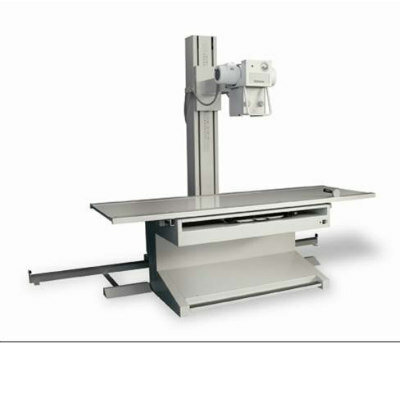
Image: The FDR Go PLUS version portable DR system (Photo courtesy of FUJIFILM).
FUJIFILM Medical Systems U.S.A, Inc., (Stamford, CT, USA), a provider of diagnostic imaging products and medical informatics solutions, displayed its suite of mobile digital radiography (DR) solutions, including its all-new FDR Go PLUS version portable DR system and the FDR AQRO portable digital X-ray system, at the 103rd scientific assembly and annual meeting of the Radiological Society of North America (RSNA).
Fujifilm’s range of diagnostic imaging products and medical informatics solutions includes digital X-ray systems, the Synapse brand of PACS, RIS and cardiovascular products, and advanced women's health imaging systems.
On display at RSNA 2017 was Fujifilm’s FDR Go PLUS version, which continues with its signature smooth, quiet travel and compact tube head, but now features a collapsible column for maximum visibility while traveling and an extra-large display for optimal previewing at the bedside, along with a new sleek redesign. Other enhancements include user adjustable drive handle (optional), wireless barcode reader (optional), an RFID card reader (optional) and extensive dedicated storage areas. Its software features include new workstation software with automated keypad display, quick start, the company’s latest Virtual Grid simulation software (optional) and Dynamic Visualization II image processing (optional).
"The nature of the radiology department demands a mobile workplace with smaller and lightweight detectors along with compact portable digital X-rays," said Johann Fernando, Ph.D., Chief Operating Officer of FUJIFILM Medical Systems U.S.A, Inc. "The FDR Go PLUS version is poised to dramatically improve workflow and performance to meet the most challenging mobile demands of imaging."
Also on display at RSNA 2017 was Fujifilm’s FDR AQRO, a complete mini-digital X-ray system, which generates high resolution images with low patient dose by combining the high sensitivity of the company’s D-EVO II detectors and its refined image processing advancements. The system provides reduced dose with Irradiation Side Sampling (ISS), improved image contrast with Virtual Grid and optimized images with Dynamic Visualization II image processing for all patient sizes and anatomy.
Latest RSNA 2017 News
- Siemens Healthineers Launches New Mammography System at RSNA
- New Real-Time Imaging AI Platform Unveiled
- Bracco Diagnostics Highlights Advancement in Diagnostic Imaging Portfolio
- Varex Imaging Showcases New X-ray Components at Trade Fair
- Samsung Unveils New Mobile CT at RSNA 2017
- Thales Unveils World’s First Portable Detector with Embedded Patient ID
- PACSHealth Showcases DoseMonitor Upgrade at Chicago Trade Show
- Carestream Exhibits New Diagnostic Imaging Solutions in Chicago
- Siemens Healthineers Spotlights Advances in Breast Imaging
- Agfa HealthCare Presents Advances in AI and Machine Learning at RSNA 2017
- Philips Showcases New Imaging Systems and Informatics Portfolio at RSNA
- Canon Showcases New DR Systems at Medical Imaging Fair
- Aspect Imaging Presents Neonatal MRI System at RSNA 2017
- Visage Imaging Debuts AI Offerings at Chicago Show
- Carestream Joins Zebra Medical Vision to Provide Access to AI Algorithms
Channels
Radiography
view channel
Novel Breast Imaging System Proves As Effective As Mammography
Breast cancer remains the most frequently diagnosed cancer among women. It is projected that one in eight women will be diagnosed with breast cancer during her lifetime, and one in 42 women who turn 50... Read more
AI Assistance Improves Breast-Cancer Screening by Reducing False Positives
Radiologists typically detect one case of cancer for every 200 mammograms reviewed. However, these evaluations often result in false positives, leading to unnecessary patient recalls for additional testing,... Read moreMRI
view channel
PET/MRI Improves Diagnostic Accuracy for Prostate Cancer Patients
The Prostate Imaging Reporting and Data System (PI-RADS) is a five-point scale to assess potential prostate cancer in MR images. PI-RADS category 3 which offers an unclear suggestion of clinically significant... Read more
Next Generation MR-Guided Focused Ultrasound Ushers In Future of Incisionless Neurosurgery
Essential tremor, often called familial, idiopathic, or benign tremor, leads to uncontrollable shaking that significantly affects a person’s life. When traditional medications do not alleviate symptoms,... Read more
Two-Part MRI Scan Detects Prostate Cancer More Quickly without Compromising Diagnostic Quality
Prostate cancer ranks as the most prevalent cancer among men. Over the last decade, the introduction of MRI scans has significantly transformed the diagnosis process, marking the most substantial advancement... Read moreUltrasound
view channel
Deep Learning Advances Super-Resolution Ultrasound Imaging
Ultrasound localization microscopy (ULM) is an advanced imaging technique that offers high-resolution visualization of microvascular structures. It employs microbubbles, FDA-approved contrast agents, injected... Read more
Novel Ultrasound-Launched Targeted Nanoparticle Eliminates Biofilm and Bacterial Infection
Biofilms, formed by bacteria aggregating into dense communities for protection against harsh environmental conditions, are a significant contributor to various infectious diseases. Biofilms frequently... Read moreNuclear Medicine
view channel
New SPECT/CT Technique Could Change Imaging Practices and Increase Patient Access
The development of lead-212 (212Pb)-PSMA–based targeted alpha therapy (TAT) is garnering significant interest in treating patients with metastatic castration-resistant prostate cancer. The imaging of 212Pb,... Read moreNew Radiotheranostic System Detects and Treats Ovarian Cancer Noninvasively
Ovarian cancer is the most lethal gynecological cancer, with less than a 30% five-year survival rate for those diagnosed in late stages. Despite surgery and platinum-based chemotherapy being the standard... Read more
AI System Automatically and Reliably Detects Cardiac Amyloidosis Using Scintigraphy Imaging
Cardiac amyloidosis, a condition characterized by the buildup of abnormal protein deposits (amyloids) in the heart muscle, severely affects heart function and can lead to heart failure or death without... Read moreGeneral/Advanced Imaging
view channel
New AI Method Captures Uncertainty in Medical Images
In the field of biomedicine, segmentation is the process of annotating pixels from an important structure in medical images, such as organs or cells. Artificial Intelligence (AI) models are utilized to... Read more.jpg)
CT Coronary Angiography Reduces Need for Invasive Tests to Diagnose Coronary Artery Disease
Coronary artery disease (CAD), one of the leading causes of death worldwide, involves the narrowing of coronary arteries due to atherosclerosis, resulting in insufficient blood flow to the heart muscle.... Read more
Novel Blood Test Could Reduce Need for PET Imaging of Patients with Alzheimer’s
Alzheimer's disease (AD), a condition marked by cognitive decline and the presence of beta-amyloid (Aβ) plaques and neurofibrillary tangles in the brain, poses diagnostic challenges. Amyloid positron emission... Read more.jpg)
CT-Based Deep Learning Algorithm Accurately Differentiates Benign From Malignant Vertebral Fractures
The rise in the aging population is expected to result in a corresponding increase in the prevalence of vertebral fractures which can cause back pain or neurologic compromise, leading to impaired function... Read moreImaging IT
view channel
New Google Cloud Medical Imaging Suite Makes Imaging Healthcare Data More Accessible
Medical imaging is a critical tool used to diagnose patients, and there are billions of medical images scanned globally each year. Imaging data accounts for about 90% of all healthcare data1 and, until... Read more
Global AI in Medical Diagnostics Market to Be Driven by Demand for Image Recognition in Radiology
The global artificial intelligence (AI) in medical diagnostics market is expanding with early disease detection being one of its key applications and image recognition becoming a compelling consumer proposition... Read moreIndustry News
view channel
Bayer and Google Partner on New AI Product for Radiologists
Medical imaging data comprises around 90% of all healthcare data, and it is a highly complex and rich clinical data modality and serves as a vital tool for diagnosing patients. Each year, billions of medical... Read more


