


Optical Scintillation Detector Monitors Radiation Treatments
|
By MedImaging International staff writers Posted on 15 Nov 2017 |
|
.")
Image: An optical system measures and validates radiation doses (Photo courtesy of RadiaDyne).
A new system provides diagnostic pinpoint accuracy measurement of the radiation treatment plan by verifying the dose delivered is consistent with the prescribed dose.
The RadiaDyne (Houston, TX, USA) OARtrac system is a real-time in vivo dosimetry device system that uses disposable scintillating detectors during stereotactic body radiotherapy (SBRT) and intensity-modulated radiation therapy (IMRT) for prostate cancer treatment or brachytherapy. The system is based on plastic scintillation detector (PSD) probes connected to a duplex fiber-optical cable that transmits the PSD signals to a charge coupled device (CCD) camera that measures the scintillator output signal.
The optical measurement is converted to an electrical signal and displayed on a digital readout located in the control room of the linear accelerator treatment machine. Based on the continuous review of the accumulative dose data provided by the system, radiation oncologists can adjust subsequent treatment if and when required, thus allowing for a true adaptive radiation therapy protocol. The PSD sensor cable can be used up to five times on the same patient.
“Radiation oncologists can now monitor multiple radiation delivery modalities within the same treatment center, as well as reduce overall treatment costs related to routine patient dose monitoring,” said John Isham, founder of RadiaDyne.
Organs at risk (OAR) are defined as normal tissues whose radiation sensitivity may significantly influence treatment planning and/or the prescribed radiation dose.
Related Links:
RadiaDyne
Gold Member
Electrode Solution and Skin Prep
Signaspray
Gold Member
Ultrasound System
FUTUS LE
Body Array Coil
12-Channel Body Array Coil 1.5 / 3.0 T
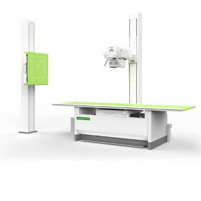
Digital Radiography System
DuraDiagnost F30

Latest Nuclear Medicine News
- New SPECT/CT Technique Could Change Imaging Practices and Increase Patient Access
- New Radiotheranostic System Detects and Treats Ovarian Cancer Noninvasively
- AI System Automatically and Reliably Detects Cardiac Amyloidosis Using Scintigraphy Imaging
- Early 30-Minute Dynamic FDG-PET Acquisition Could Halve Lung Scan Times
- New Method for Triggering and Imaging Seizures to Help Guide Epilepsy Surgery
- Radioguided Surgery Accurately Detects and Removes Metastatic Lymph Nodes in Prostate Cancer Patients
- New PET Tracer Detects Inflammatory Arthritis Before Symptoms Appear
- Novel PET Tracer Enhances Lesion Detection in Medullary Thyroid Cancer
- Targeted Therapy Delivers Radiation Directly To Cells in Hard-To-Treat Cancers
- New PET Tracer Noninvasively Identifies Cancer Gene Mutation for More Precise Diagnosis
- Algorithm Predicts Prostate Cancer Recurrence in Patients Treated by Radiation Therapy
- Novel PET Imaging Tracer Noninvasively Identifies Cancer Gene Mutation for More Precise Diagnosis
- Ultrafast Laser Technology to Improve Cancer Treatment
- Low-Dose Radiation Therapy Demonstrates Potential for Treatment of Heart Failure
- New PET Radiotracer Aids Early, Noninvasive Detection of Inflammatory Bowel Disease
- Combining Amino Acid PET and MRI Imaging to Help Treat Aggressive Brain Tumors
Channels
Radiography
view channel")
Novel Breast Imaging System Proves As Effective As Mammography
Breast cancer remains the most frequently diagnosed cancer among women. It is projected that one in eight women will be diagnosed with breast cancer during her lifetime, and one in 42 women who turn 50... Read more")
AI Assistance Improves Breast-Cancer Screening by Reducing False Positives
Radiologists typically detect one case of cancer for every 200 mammograms reviewed. However, these evaluations often result in false positives, leading to unnecessary patient recalls for additional testing,... Read more")
")
MRI
view channel")
PET/MRI Improves Diagnostic Accuracy for Prostate Cancer Patients
The Prostate Imaging Reporting and Data System (PI-RADS) is a five-point scale to assess potential prostate cancer in MR images. PI-RADS category 3 which offers an unclear suggestion of clinically significant... Read more")
Next Generation MR-Guided Focused Ultrasound Ushers In Future of Incisionless Neurosurgery
Essential tremor, often called familial, idiopathic, or benign tremor, leads to uncontrollable shaking that significantly affects a person’s life. When traditional medications do not alleviate symptoms,... Read more")
Two-Part MRI Scan Detects Prostate Cancer More Quickly without Compromising Diagnostic Quality
Prostate cancer ranks as the most prevalent cancer among men. Over the last decade, the introduction of MRI scans has significantly transformed the diagnosis process, marking the most substantial advancement... Read more of magnetic field vs. 1.5 and 3 T for conventional MRI machines in hospitals (Photo courtesy of CEA)")
Ultrasound
view channel")
Deep Learning Advances Super-Resolution Ultrasound Imaging
Ultrasound localization microscopy (ULM) is an advanced imaging technique that offers high-resolution visualization of microvascular structures. It employs microbubbles, FDA-approved contrast agents, injected... Read more")
Novel Ultrasound-Launched Targeted Nanoparticle Eliminates Biofilm and Bacterial Infection
Biofilms, formed by bacteria aggregating into dense communities for protection against harsh environmental conditions, are a significant contributor to various infectious diseases. Biofilms frequently... Read more")
")
General/Advanced Imaging
view channel")
New AI Method Captures Uncertainty in Medical Images
In the field of biomedicine, segmentation is the process of annotating pixels from an important structure in medical images, such as organs or cells. Artificial Intelligence (AI) models are utilized to... Read more.jpg "Image: CT coronary angiography for diagnosing coronary artery disease offers advantages over other diagnostic tests (Photo courtesy of 123RF)")
CT Coronary Angiography Reduces Need for Invasive Tests to Diagnose Coronary Artery Disease
Coronary artery disease (CAD), one of the leading causes of death worldwide, involves the narrowing of coronary arteries due to atherosclerosis, resulting in insufficient blood flow to the heart muscle.... Read more")
Novel Blood Test Could Reduce Need for PET Imaging of Patients with Alzheimer’s
Alzheimer's disease (AD), a condition marked by cognitive decline and the presence of beta-amyloid (Aβ) plaques and neurofibrillary tangles in the brain, poses diagnostic challenges. Amyloid positron emission... Read more.jpg "Image: Exemplary illustration of the labeling and segmentation process (Photo courtesy of TUM)")
CT-Based Deep Learning Algorithm Accurately Differentiates Benign From Malignant Vertebral Fractures
The rise in the aging population is expected to result in a corresponding increase in the prevalence of vertebral fractures which can cause back pain or neurologic compromise, leading to impaired function... Read moreImaging IT
view channel")
New Google Cloud Medical Imaging Suite Makes Imaging Healthcare Data More Accessible
Medical imaging is a critical tool used to diagnose patients, and there are billions of medical images scanned globally each year. Imaging data accounts for about 90% of all healthcare data1 and, until... Read more
Global AI in Medical Diagnostics Market to Be Driven by Demand for Image Recognition in Radiology
The global artificial intelligence (AI) in medical diagnostics market is expanding with early disease detection being one of its key applications and image recognition becoming a compelling consumer proposition... Read more

Industry News
view channel")
Bayer and Google Partner on New AI Product for Radiologists
Medical imaging data comprises around 90% of all healthcare data, and it is a highly complex and rich clinical data modality and serves as a vital tool for diagnosing patients. Each year, billions of medical... Read more")
")
")